
Corneal Cross Linking
Corneal Cross Linking
Corneal Crosslinking
Corneal crosslinking is an advanced procedure offered at Focus Eye Care, serving patients in Queens, NYC, and Long Island. This minimally invasive treatment aims to strengthen the cornea, helping to slow the progression of conditions like keratoconus and corneal ectasia.
Our experienced team is dedicated to providing personalized care and guiding you through your treatment options, ensuring you receive the support you need to protect and preserve your vision.

Corneal crosslinking, sometimes referred to as CXL or corneal collagen cross-linking, is a minimally invasive procedure designed to treat progressive keratoconus and post-surgical ectasia. Historically, patients with keratoconus or ectasia had limited options, relying on rigid contact lenses or, in more severe cases, corneal transplants. However, advances in corneal crosslinking have transformed the management of these conditions.
Statistics show that without treatment, up to 1 in 5 individuals with keratoconus may eventually require a corneal transplant.1 Corneal crosslinking works by strengthening the cornea, which has been compromised by conditions like keratoconus or prior refractive surgery. This procedure aims to stabilize the cornea, slowing or halting disease progression and thereby reducing the risk of vision loss. In some cases, corneal crosslinking can also enhance visual acuity.
What Does Corneal Crosslinking Treat?
Corneal crosslinking, sometimes referred to as “keratoconus surgery,” has revolutionized the treatment of keratoconus, offering a more effective approach to managing this progressive eye condition. In addition to keratoconus, corneal crosslinking can also be used to treat corneal ectasia, and researchers are investigating its potential for other corneal diseases.
Corneal Crosslinking for Keratoconus
Keratoconus is a condition in which the cornea, the transparent front layer of the eye, gradually thins and bulges into a cone-like shape. This change in shape leads to distorted vision, which worsens as the disease progresses. Corneal crosslinking aims to halt the progression of keratoconus by strengthening the cornea, helping to preserve vision and prevent further deterioration.2
Corneal Crosslinking for Corneal Ectasia
Corneal ectasia is a condition characterized by a thinning or weakening of the cornea. It can occur in various forms, such as keratoconus, keratoglobus, pellucid marginal degeneration (PMD), and terrien marginal degeneration (TMD). In rare cases, corneal ectasia can develop as a complication following refractive surgeries like LASIK or PRK. Corneal crosslinking provides a treatment option to stabilize the cornea and reduce the risk of vision loss in patients with ectasia after vision correction surgery.
How Does Corneal Crosslinking Work?
Corneal crosslinking is designed to stop the progression of keratoconus and corneal ectasia by strengthening the collagen fibers within the cornea. This minimally invasive procedure uses riboflavin (Vitamin B2) eye drops in combination with ultraviolet light (UV) therapy to enhance the cornea’s stability and reduce the risk of further damage.
During the procedure, riboflavin eye drops are applied to the cornea. Once the cornea is sufficiently saturated with riboflavin, it is exposed to UV light. The interaction between the riboflavin and UV light creates new cross-links between the collagen fibers, increasing the cornea’s strength and rigidity, thereby helping to preserve vision.

Preparing for Corneal Crosslinking
The first step in preparing for corneal crosslinking is to schedule a consultation with a qualified ophthalmologist. During this appointment, your eye doctor will conduct a thorough eye exam to confirm your diagnosis and determine if you are a suitable candidate for the procedure. This is also an excellent opportunity to ask any questions you may have about the treatment process and what to expect.
If corneal crosslinking is right for you, our team will provide you with detailed pre-procedure instructions to ensure the best possible outcome. It’s important to plan for your recovery as well. Since you won’t be able to drive yourself home after the procedure, please arrange for transportation in advance.
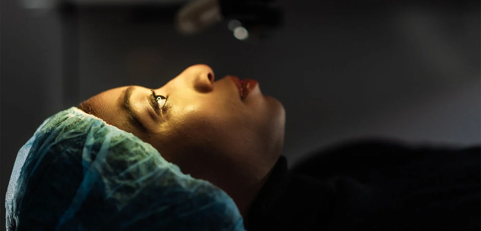
The Corneal Crosslinking Procedure
Corneal crosslinking is typically performed as an outpatient procedure and usually takes about an hour, although you should plan to be at our eye care center for a few hours on the day of your treatment. Before the CXL procedure begins, anesthetic eye drops are applied to numb the eyes, ensuring you remain comfortable throughout. In some cases, an oral sedative may be provided to help you relax.
During the procedure, the thin outer layer of the cornea, known as the epithelium, is carefully removed to allow the riboflavin eye drops to penetrate the corneal tissue. These drops are applied for approximately 30 minutes. In the final stage, the cornea is exposed to UV light for an additional 30 minutes, which helps strengthen the cornea by forming new cross-links between the collagen fibers.
Once the treatment is complete, a bandage contact lens is placed on the eye to protect it during the healing process. This lens typically stays in place for about a week while the eye recovers.
Recovery After Corneal Crosslinking
After corneal crosslinking, a bandage contact lens will be placed on the treated eye to protect it during the initial healing period. As the numbing drops wear off, you may experience some discomfort, such as a burning sensation, grittiness, or increased sensitivity to light. This is normal and should subside as your eye heals.
You will be prescribed antibiotic eye drops to use for about a week to prevent infection, along with steroid eye drops for a few weeks to help reduce inflammation. During the first week, it’s important to avoid water, sweat, and makeup around the eyes to prevent irritation and ensure proper healing. Refrain from rubbing your eyes for at least the first five days to avoid disrupting the healing process.
Plan to have a friend or family member drive you home after the procedure, as your vision may be blurred and your eyes sensitive. It’s advisable to take a few days off work to allow your eyes to recover comfortably. Our team will provide you with detailed post-operative care instructions to support a smooth recovery.
FAQs about Corneal Cross Linking
If you have been diagnosed with keratoconus or corneal ectasia, corneal crosslinking may be an option worth exploring. Ideal candidates are typically over the age of 14 and have mild to moderate stages of keratoconus. A comprehensive eye exam and consultation with your ophthalmologist will determine if this procedure is right for you.
No, if treatment is needed in both eyes, they will be addressed on separate days. This approach allows one eye to heal properly before proceeding with the second treatment.
Yes, clinical studies have shown that corneal crosslinking can significantly improve symptoms of keratoconus and slow its progression.3 Patients who undergo the procedure often experience a stabilization of their vision, reducing the likelihood of further vision loss.
No, the appearance of your eyes will not change after corneal crosslinking. The treatment works to strengthen the cornea internally, so there is no visible difference in the way your eyes look.
Many insurance companies provide coverage for corneal crosslinking. We work with a variety of insurance plans and also offer financing options to make this treatment accessible. Contact our office to learn more about payment options.
The procedure itself should not be painful due to the use of numbing eye drops. However, some discomfort is common during the recovery period. Most patients find relief with over-the-counter pain medications, but prescription options are available if needed.


Are You a Candidate for
Vision Correction?
Whether you just need a general eye exam, or if you are interested in reducing or eliminating your need for glasses or contacts, our team is here to help! Contact us to book an appointment today!


1 Pramanik S, Musch DC, Sutphin JE, Farjo AA. Extended long-term outcomes of penetrating keratoplasty for keratoconus. Ophthalmology. 2006 Sep;113(9):1633-8. doi: 10.1016/j.ophtha.2006.02.058. Epub 2006 Jul 7. PMID: 16828503. Available: https://pubmed.ncbi.nlm.nih.gov/16828503/. Accessed September 17, 2024.
2 American Academy of Ophthalmologists. What is Keratoconus? Available: https://www.aao.org/eye-health/diseases/what-is-keratoconus. Accessed September 17, 2024.
3 Belin MW, Lim L, Rajpal RK, Hafezi F, Gomes JAP, Cochener B. Corneal Cross-Linking: Current USA Status: Report From the Cornea Society. Cornea. 2018 Oct;37(10):1218-1225. doi: 10.1097/ICO.0000000000001707. Erratum in: Cornea. 2019 Oct;38(10):e49. doi: 10.1097/ICO.0000000000001901. PMID: 30067537. Available: https://pubmed.ncbi.nlm.nih.gov/30067537/. Accessed September 17, 2024.
The doctors at Focus Eye Care & Surgery have reviewed and approved this content.
Page Updated: