Conditions We Treat
at Focus Eye Care & Surgery
Our сlinic offers a wide range of private ophthalmologist services.
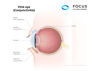
Pink Eye
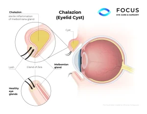
Chalazion
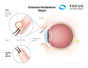
Stye
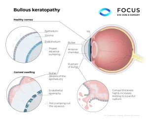
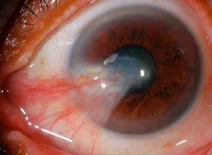
Bullous keratopathy
Pterygium
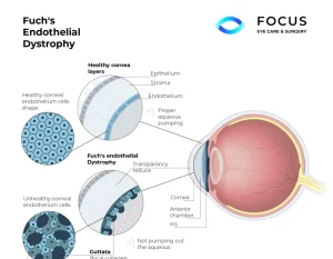
Fuchs Endothelial Dystrophy
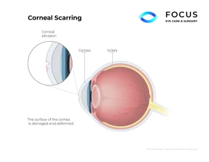
Corneal Scarring
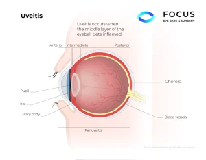
Uveitis
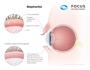
Blepharitis
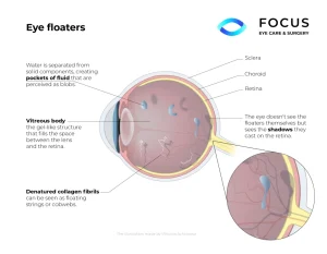
Eye Floaters Eye Flashes
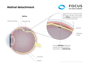
Retinal Detachment
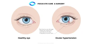
Ocular Hypertension
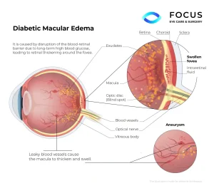
Diabetic macular edema
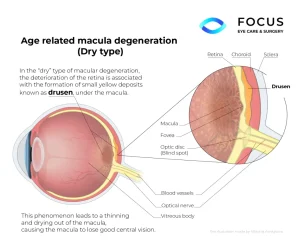
Age related macular degeneration
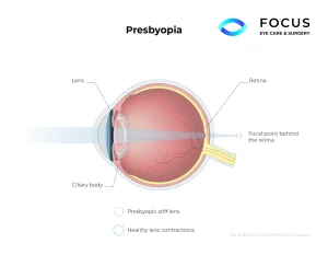
Presbyopia
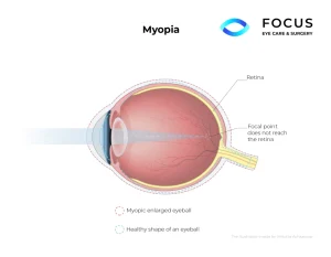
Myopia
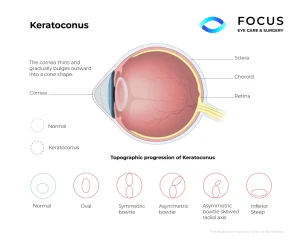
Keratoconus
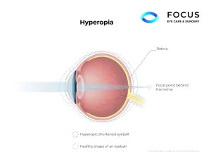
Hyperopia
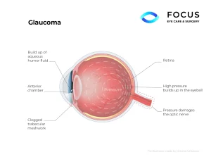
Glaucoma
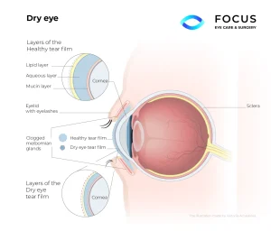
Dry Eye
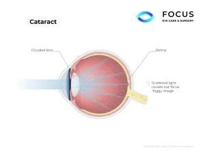
Cataracts
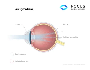
Astigmatism